Figure 6.
Histologic Characteristics
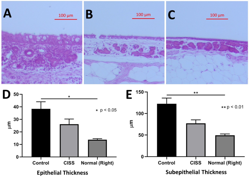
A. H&E staining of maxillary sinus at week 4 with non-drug eluting (control) stent (x10, Scale bar = 100μm)
B. H&E staining of maxillary sinus at week 4 with CISS (x10, Scale bar = 100μm)
C. H&E staining of maxillary sinus at week 4 on right normal side (x10, Scale bar = 100μm)
D. The height of the epithelial cell layer: Sinus epithelia from those rabbits treated with non-drug eluting stents were the thickest (38.31 +/− 5.7 μm), followed by CISS (26.07 +/− 4.2 μm) and normal controls (13.75 +/− 0.80 μm) (Welch’s ANOVA test, * p = 0.02)
E. The thickness of the submucosal layer: Submucosal layer was also the thickest in those sinusitis rabbits who had non-drug eluting (control) stents (122.6 +/− 13.16 μm), followed by sinusitis rabbits treated with CISS (77.08 +/− 8.23 μm) and placebo (49.60 +/− 3.0 μm) (Welch’s ANOVA, ** p = 0.008)
CISS: Ciprofloxacin Ivacaftor coated Sinus Stent

