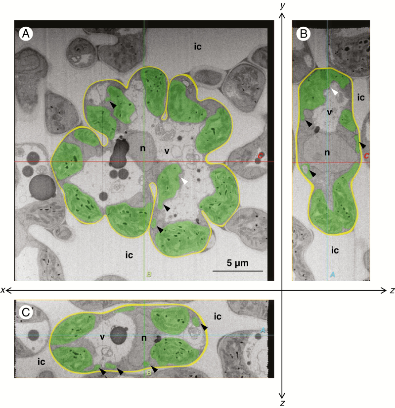
Fig. 2.
Central cross-sections of the mesophyll cells of the rice leaf blade in the NaCl-treated plants. (A) xy-transversal sections obtained with the focused ion beam scanning electron microscope. One image in a sequence of sections for a whole mesophyll cell: 84th in the sequence of 154. (B, C) Orthogonal slice images virtually imaged with the plugin ‘Volume Viewer’ of the software ‘Fiji’. (B) yz-orthogonal sections of the image stacks. (C) xz-orthogonal sections of the image stacks. Each slice plane (A, B and C) crosses the lines marked ‘A, B and C’ shown in the other orientational images. All images were segmented manually: green, chloroplast; yellow, cell wall; ic, intercellular space; n, nucleus; v, vacuole. White arrowheads indicate the partly distorted portion of the chloroplast envelope. Black arrowheads indicate the chloroplast protrusions or vesicles. Cutting interval (z-step) = 50 nm.

