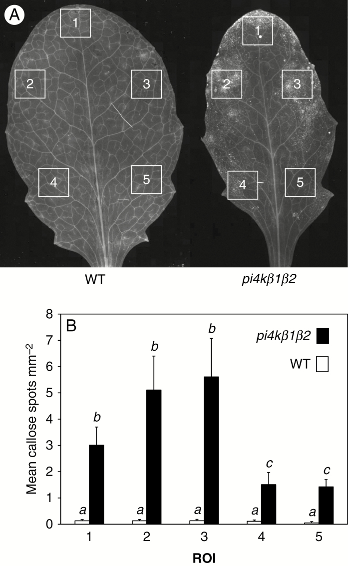
Fig. 3.
Pattern of callose accumulation in pi4kβ1β2 leaves. (A) Aniline blue staining, and fluorescence microscopy. Scale bar = 500 µm. (B) Callose particles accumulated in different ROI. The squares represent the ROI. Data are presented as means ±s.e.m. Statistical differences were assessed using a two-way ANOVA, with a Tukey honestly significant difference (HSD) multiple mean comparison post hoc test. Different letters indicate a significant difference, Tukey HSD, P < 0.05. n = 11.

