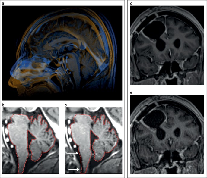
Fig. 3.
Patient positioning for simulation MRI. a Simulation MRI with mask immobilization vs. in a diagnostic head coil. The head is substantially flexed in the diagnostic head coil (amber) compared to the simulation MRI with mask immobilization (blue) leading to slight displacement of infratentorial structures (b, c, white arrows). d, e Reduced motion artifacts in the stereotactic mask system (d) compared to the diagnostic head coil (e)

