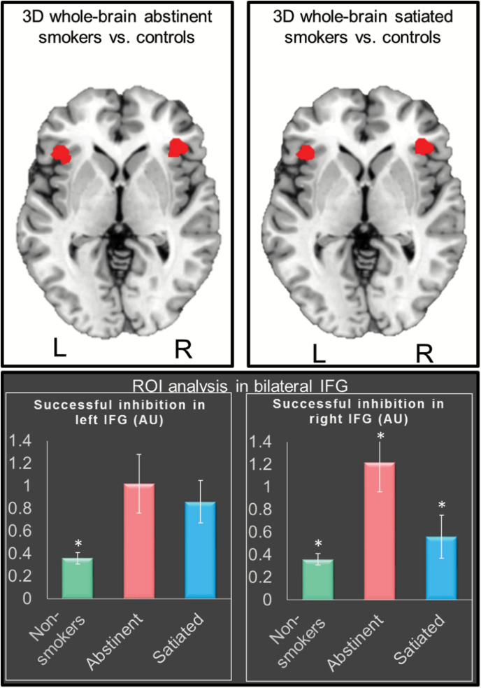
Figure 1.
Top row: Whole-brain analyses showing greater successful inhibition activity (t maps cluster-corrected at p < .05, cluster size ≥ 421) in abstinent smokers and satiated smokers relative to controls in bilateral inferior frontal gyrus (IFG), with 81% overlap across the two comparisons. Bottom row: Region of interest (ROI)-level analyses in the bilateral IFG showing higher successful inhibition activity in abstinent smokers relative to satiated and nonsmokers. The ROIs were defined as the overlapping areas across the two whole-brain comparisons. *Significantly different from the two other groups with p < .001.

