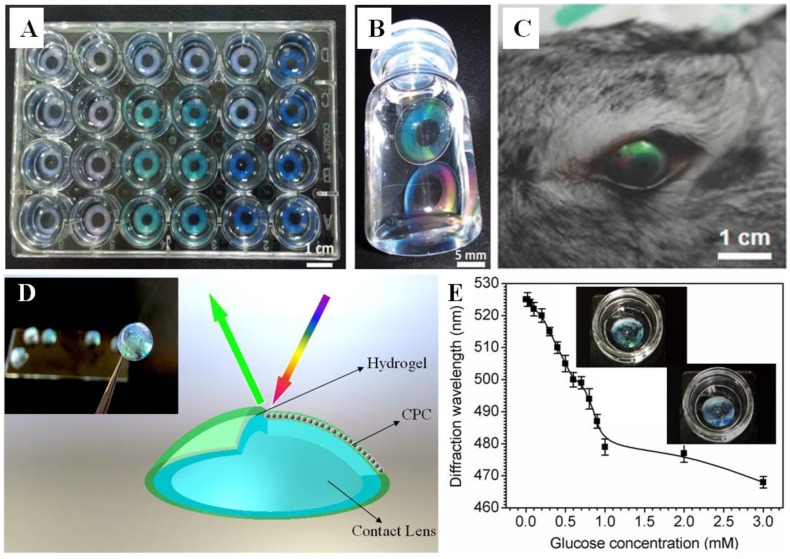
Figure 6.
(A,B) Photographs of structural colored PHEMA contact lenses [88]. (C) Photograph of a rabbit wearing a structurally colored contact lens sensor [92]. (D) Diagram and photograph (insert) of a HCPC contact lens [6]. (E) The response of a PVA HCPC contact lens at low glucose concentration; insert shows a photograph of the color changed lens sample [93].

