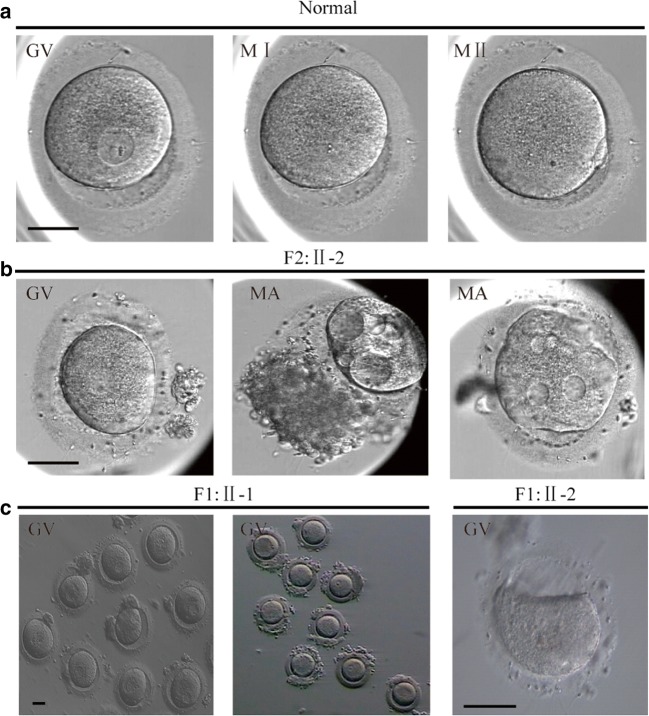
Fig. 1.
Phenotypes of oocytes from patients and fertile control. a Morphology of GV, MI, and MII oocytes from a fertile control individual. b Oocytes arrested at the GV stage and morphologic abnormalities from II-2 (F2). c GV stage oocytes from II-1 and II-2 (F1). Scale bar, 50 μm. F2, Family2; F1, Family1; GV, Germinal Vesicle; MI, Metaphase I; MII, Metaphase II; MA, morphology abnormality

