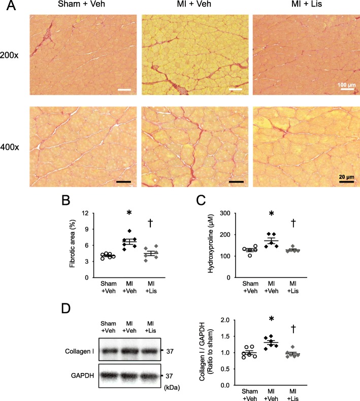
Fig. 3.
Effects of Lis administration on skeletal muscle fibrosis in MI mice. a Representative high-power photomicrographs of gastrocnemius cross-sections stained with picrosirius red from Sham, MI, and MI + Lis mice at 14 days postsurgery. Top panels show low magnification micrographs (× 200; white scale bars = 100 μm), and bottom panels show higher magnification micrographs (× 400; black scale bars = 20 μm). b Summary data of fibrotic area (n = 6). c Quantification of hydroxyproline levels in soleus muscle from 3 groups at 14 days postsurgery (n = 5). d Representative data of immunoblotting of gastrocnemius muscle lysates obtained from the 3 groups (left). Right: summarized data of collagen protein expression level (n = 6). GAPDH (glyceraldehyde-3-phosphate dehydrogenase) was used as an internal control. The sizes of the molecular weight markers are indicated on the right in kilo Dalton. Data are expressed as the mean ± SE. *P < 0.05 vs. Sham + Veh, †P < 0.05 vs. MI + Veh

