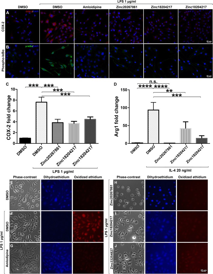
Fig. 5.
COX-2, phopho-IκBα, and reactive oxygen species expression in LPS-stimulated BV-2 cells with and without L-VGCC blockade. Effects of L-VGCC blockade on COX-2 expression (a) and detection of phosphorylated IκBα (b) in BV-2 cells stimulated with LPS. Quantitative RT-PCR analysis of COX-2 expression in BV-2 cells treated with DMSO control (2 μl/ml), Zinc20267861 (2.0 μM), Zinc18204217 (2.0 μM), and Zinc33254827 (2.5 μM) stimulated with LPS 1 μg/ml (c). Arg-1 expression with the same treatments in BV-2 cells stimulated with IL-4 (20 ng/ml) (d) (n = 3 with 3 technical replicates). A one-way ANOVA test with Tukey multiple comparisons was used to determine statistical significance. p > 0.05; ***p < 0.001. Target genes were normalized to cyclophilin and presented as the fold change of DMSO control. Detection of reactive oxygen species in BV-2 cell culture, minimal oxidation of dihydroethidium in DMSO 2 μl/ml untreated control cells (e); strong oxidized ethidium signals detected when stimulated with LPS 1 μg/ml (e). Treatment with 3.5 μM of amlodipine (g), Zinc20267861 (2.0 μM) (h), Zinc18204217 (2.0 μM) (i), and Zinc33254827 (2.5 μM) (j) demonstrated marked reduction of the oxidized ethidium signals in BV-2 cell culture.

