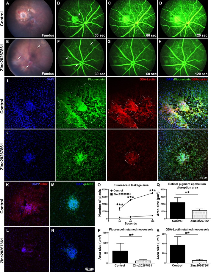
Fig. 6.
Effects of L-VGCC blockade on laser-induced CNV and expression of COX-2 and phospho-Iκbα in mice. Fundus in vivo images of vehicle-treated laser CNV fundus 5 days after induction of the model (a) and Zinc20267861 (10 μg) subconjunctival treated eyes (e). Arrows indicate laser burn spots. In vivo fluorescent angiography in vehicle-treated laser CNV animals 30 (b), 60 (c), and 120 (d) after subcutaneous injection of fluorescein sodium 1 mg/kg and same time points (f–h) for Zinc20267861 (10 μg) subconjunctival treated eyes. Arrows indicate fluorescein leakage areas. Ex vivo evaluation of the laser spot sizes in retinal pigment epithelia choroid-scleral complexes (RCSC) flat mounts in control (i) and Zinc20267861 (10 μg) treated eyes (j) counterstained with GSA-lectin. Immunofluorescent detection of COX-2 (k, l) and phospho-Iκbα (m, n) in RCSC flat mounts in control and Zinc20267861-treated eyes. Quantitative analysis of fluorescein leakage area over time in vivo (o). A one-way ANOVA test with Tukey multiple comparisons was used to determine statistical significance. ***p < 0.001; (n = 3). Ex vivo quantitative analysis of fluorescein-stained neovessels (n = 6) (p), retinal pigment epithelia disruption area (n = 6) (q), and GSA-lectin stained neovessels (n = 6) (r). Student t-test was used to determine statistical significance between two groups. *p < 0.05; **p < 0.01; ***p < 0.001

