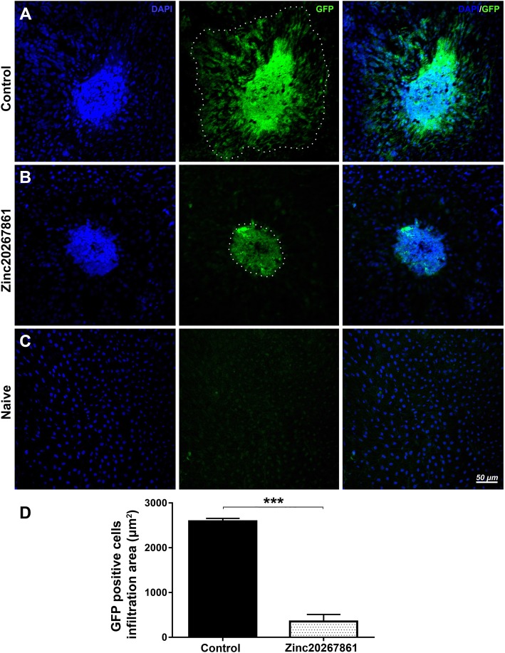
Fig. 7.
GFP-positive microglia cells and monocytes infiltration of laser CNV spot ex vivo in CX3CR1gfp/+ mice with L-VGCC blockade. Retinal pigment epithelia choroid-scleral complexes (RCSC) flat mounts indicate infiltration with GPF-positive cells of the laser CNV spot in vehicle-treated control CX3CR1gfp/+ animals (a) or Zinc20267861 (10 μg) (b) subconjunctivally treated eyes 5 days after model induction. Naive control CX3CR1gfp/+ mice RCSC flat mounts shows minimal presence of GFP-positive cells (c). Quantitative analysis of GFP-positive cell infiltration area (d). Student t-test was used to determine statistical significance. ***p < 0.001, (n = 3)

