Fig. 5.
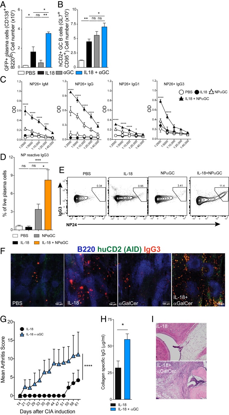
Cognate activation of iNKT cells during chronic inflammation provides B cell help and IL-18 + αGalCer exacerbates collagen-induced arthritis. (A and B) Percent of (A) GFP+ plasma cells and (B) hCD2+ germinal center B cells from spleens of Aicda Cre GT Rosa Flox mice treated as indicated. Data are representative of three experiments (n = 3 mice per group). (C) OD (450 nm) of NP-reactive IgM, IgG, IgG1, and IgG3 measured by ELISA. Sera were diluted as shown on the x axis. Data are representative of two experiments (n = 4 or 5 mice per group). (D) Percent of intracellular, NP24-reactive IgG3+ cells among plasma cells (CD138+, B220−). (E) Representative flow cytometry plots for intracellular staining. (F) Representative immunofluorescence microscopy of the spleen (day 12) showing B220 (blue), hCD2 (green), and IgG3 (red) antibody-forming cells. (Scale bars, 100 μm.) Data are representative of two experiments (n = 4 or 5 mice per group). One-way (A, B, D) or two-way (C) ANOVA, Student's t test (H). *P<0.05, **P<0.005, ***P<0.001, ****P<0.0001. Graphs display group mean ± SEM. (G) Arthritis score among B10.Q mice immunized with CII and 1 d later treated i.p. with IL-18 (2 μg) or IL-18 (2 μg) + αGalCer (5 μg); two-way ANOVA. (H) Comparison of collagen type II-specific IgG titers. (I) Examples of histological analysis of hind limbs using H&E staining.

