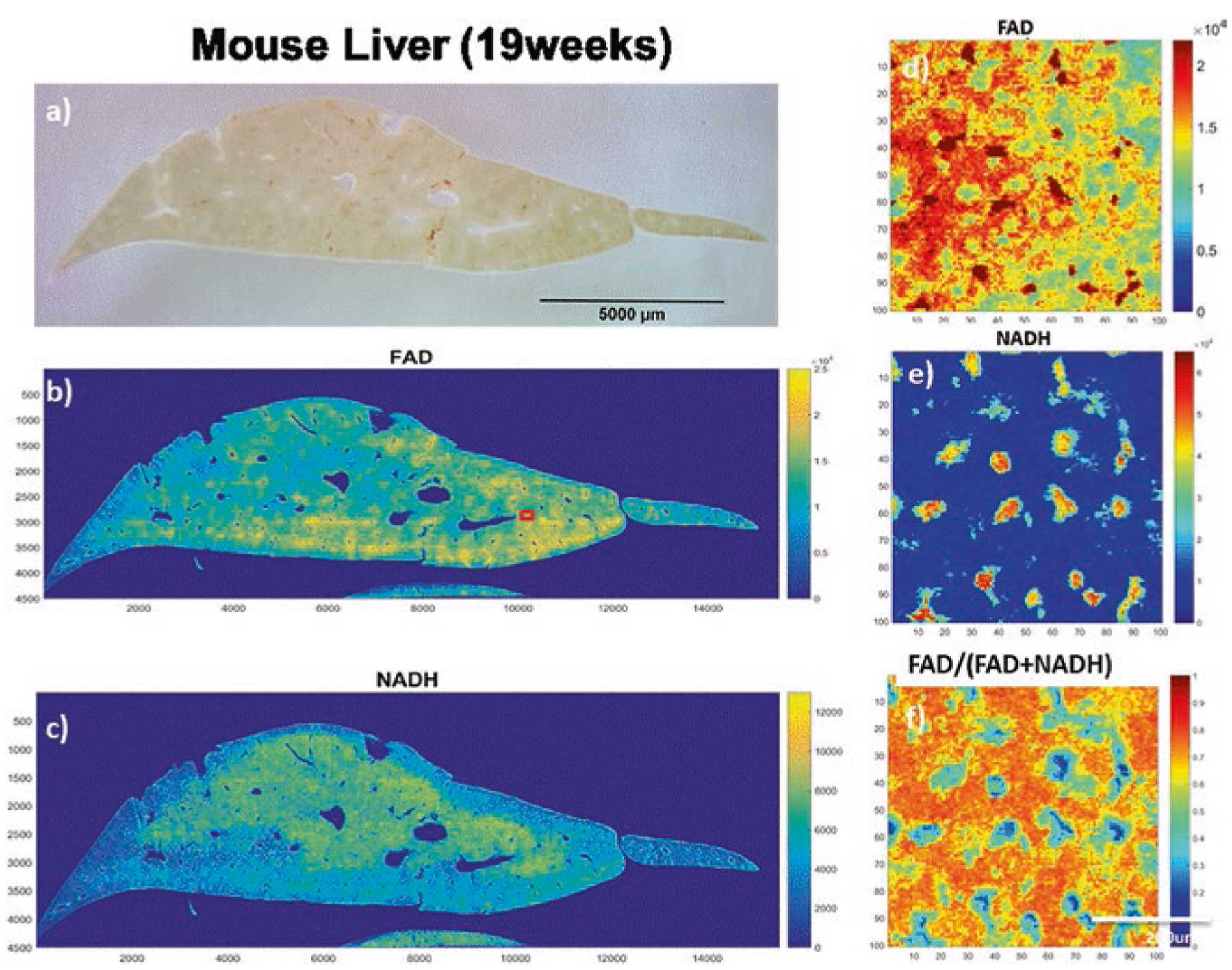
Fig. 2.

Optical redox images of a fixed mouse liver sample from the two-photon scanner. (a) White light photo of the liver sample (19 weeks); (b, c) FAD and NADH images, respectively; (d–f) Blown-up FAD, NADH and redox ratios images, respectively, for an area of interest (100 × 100 pixel) marked as a red square in (b)
