Fig. 6.
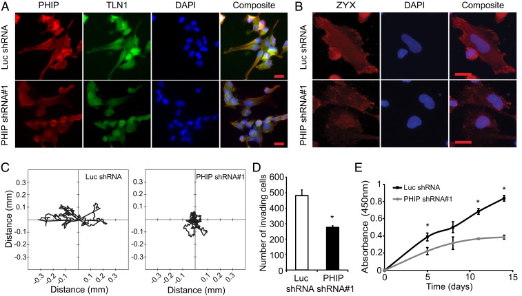
Effects of stable suppression of PHIP in 3832 primary glioblastoma transformants stably expressing anti-luc shRNA or anti-PHIP shRNA#1. (A) Qualitative immunofluorescence analysis of PHIP and TLN1 expression in primary 3832 transformants. DAPI staining was used to counterstain the nuclei. Quantification of the immunofluorescence results, including statistical analysis, is provided in SI Appendix, Fig. S8. (B) Qualitative immunofluorescence analysis of ZYX expression in primary 3832 transformants. DAPI staining was used to counterstain the nuclei. Quantification of the immunofluorescence results, including statistical analysis, is provided in SI Appendix, Fig. S8. (C) Two-dimensional projections of cell tracks from primary 3832 transformants stably expressing anti-PHIP shRNA#1 (n = 9 cells) or anti-luc shRNA (n = 11 cells) plotted normalized to their starting positions. (D) Invasion into Matrigel of primary 3832 transformants. (E) Cell survival analysis of primary 3832 transformants. All graphs represent mean ± SEM, *P < 0.05 versus control. (Scale bars: 20 µm.)

