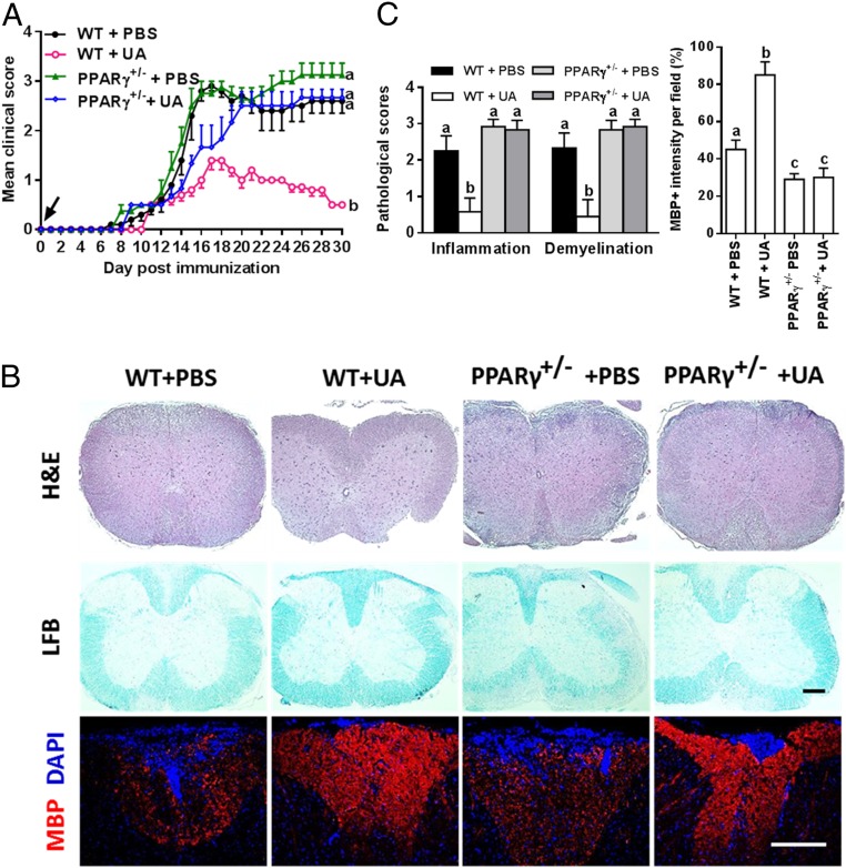
Fig. 3.
The therapeutic effect of UA on CNS autoimmunity is PPARγ dependent. (A) Clinical score of UA- or PBS-treated WT (PPARγ+/+) or PPARγ+/− mice (C57BL/6J background). (B) Sections (lumbar) were assayed for inflammation by H&E, demyelination by LFB, and MBP expression by immunostaining. Dorsal column at the thoracic spinal cord is shown as representative images. (C) CNS pathology was scored on a 0 to 3 scale, and MBP intensity was measured in the white matter of spinal cord using Image-Pro. The measured areas included 8 to 10 fields and covered virtually all of the white matter of the spinal cord. Groups designated by the same letter are not significantly different, while those with different letters (a, b, or c) are significantly different (P < 0.05–0.01). All quantifications were made from three independent experiments. Symbols represent mean ± SD; n = 5 to 8 mice per group. (Scale bar, 100 µm in B.)

