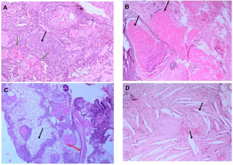
Figure 3.
Hematoxylin and Eosin (H&E) stained pictures of ACP tumor. (A) Histology of ACP, showing loosely cohesive squamous cells surrounded by peripherally palisading cells with distinct wet keratin (green arrows), stellate reticulum (black arrow); (B) an aggregate of epithelium with squamous nodule multiple wet keratin (black arrows, 20x); (C) basal epithelial layer showing a pattern of picket-fence (red arrow), and gliotic brain tissue (black arrow); (D) large number of white clefts representing cholesterol deposits (black, arrow).

