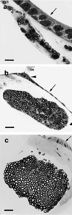
Fig. 5.
(right) Light micrographs of transverse sections of the amphibian papillar branch of the Xenopus laevis VIIIth cranial nerve. Arrows indicate lumen of the papillar epithelium. Large arrowhead indicates the attachment point of the tectorial membrane. (a) Larval stage 52, (b) juvenile, 1 day postmetamorphosis, (c) adult. Bars = (a) 10 μm, (b,c) 25 μm. tm, tectorial membrane.

