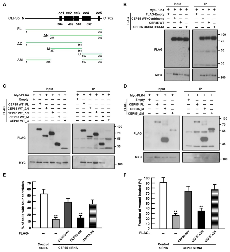
Figure 2. CEP85 interacts functionally with PLK4.
(A). Domain overview of human CEP85. cc coiled-coil. Vectors that were used in this work are shown by green lines. (B-D). Detection of expressed FLAG-CEP85 WT, Q640A+E644A mutant or fragments coimmunoprecipitating with Myc- PLK4. (E-F). U-2 OS cells expressing FLAG or the siRNA-resistant FLAG-CEP85 transgene were transfected with control or CEP85 siRNA for 72 h. (E). The graph indicates the percentage of cells with four centrioles at normal serum conditions (n = 100, three independent experiments). (F). The graph indicates the percentage of the percentage of wound area recovered at low serum conditions (n = 3/experiment, three independent experiments). Two-tailed t-test was performed for all p-values (**p < 0.01, *p <0.05).

