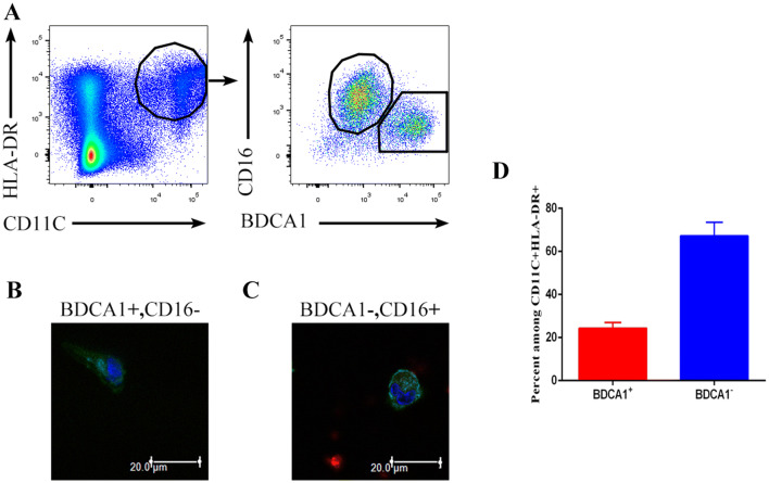
Fig. 1.
Identification of DCs in human malignant pleural effusions. a Light density cells from the malignant pleural effusions of NSCLC patients were stained with anti-HLA-DR, CD11C, CD16 and CD1c antibodies and analyzed by flow cytometry. One representative experiment out of 8 is shown. Sorted HLA-DR+ CD11c+ CD16− BDCA1+ (b) and HLA-DR+ CD11c+ CD16+ BDCA1− (c) cells from the malignant pleural effusions were analyzed by laser-scanning confocal microscopy. One representative experiment out of three is shown. d Percentage of CD16−BDCA1+ and CD16+BDCA1− cells among the CD11C+HLA-DR+ cells from the malignant pleural effusion of NSCLC patients. The mean ± SD is shown (n = 12)

