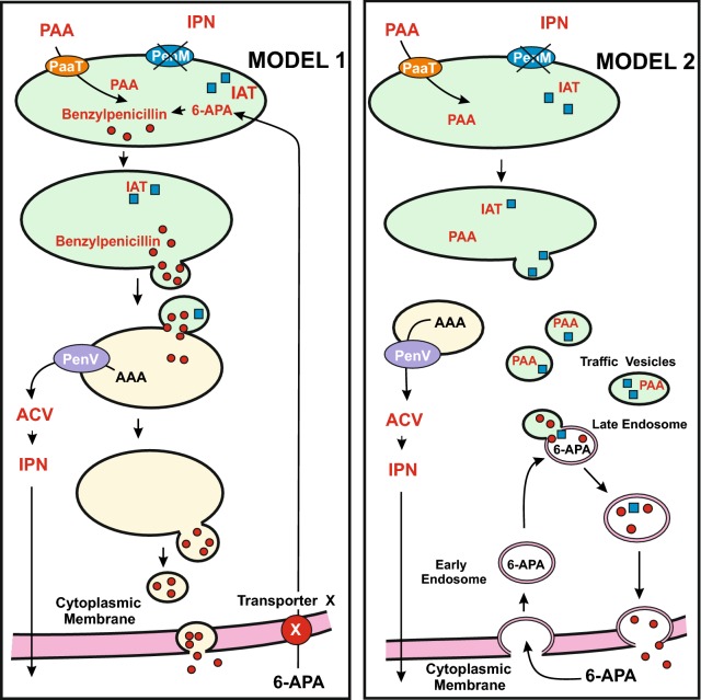
Fig. 3.
Models for the conversion of external 6-APA into benzylpenicillin and its secretion in penM mutants blocked in IPN import in peroxisomes. Peroxisomes are shown as green ellipses. Vacuoles are shown as yellow ellipses. Model 1 (left side). Conversion of 6-APA into benzylpenicillin in peroxisomes. 6-APA may be introduced in the cells through an unknown cell membrane transporter X, indicated by a red circle. The transporter PenV, indicated with a purple ellipse, exports α-aminoadipic acid from vacuoles to the cytosol. The IAT protein is shown as small blue squares. Model 2 (right side). Conversion of 6-APA into benzylpenicillin in cytosolic traffic vesicles where the acylation of 6-APA may occur. This implies that IAT is transferred to traffic vesicles. In this model 6-APA is introduced by transporters or endocytosis and is translocated by early endosomes to traffic vesicles. Secretion of benzylpenicillin through the cell membrane/cell wall occurs by fusion with the cell membrane (see text for additional information). For simplicity the early steps of the pathway (biosynthesis of ACV and IPN) are not shown

