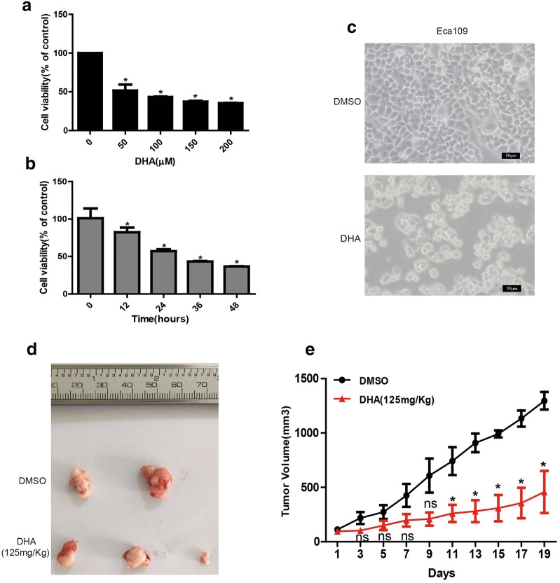
Fig. 1.
The anticancer effect of DHA on Eca109 cells in vitro and in vivo. a, b Cells were incubated with 0, 50, 100, 150, 200 μM of DHA for 48 h (a) or treated with 100 μM of DHA for 0, 12, 24, 36, 48 h (b) and then were collected for measuring the percentage of viable cells with crystal violet assay. (c) The morphological characteristics of Eca109 cells after treated with DHA (100 μM) or DMSO. (d, e) The tumor size (d) and proliferation curve (e) from esophageal cancer xenograft from mice. Data represent mean ± S.D. *p < 0.05 was significant difference between DHA-treated and control groups

