Fig. 5.
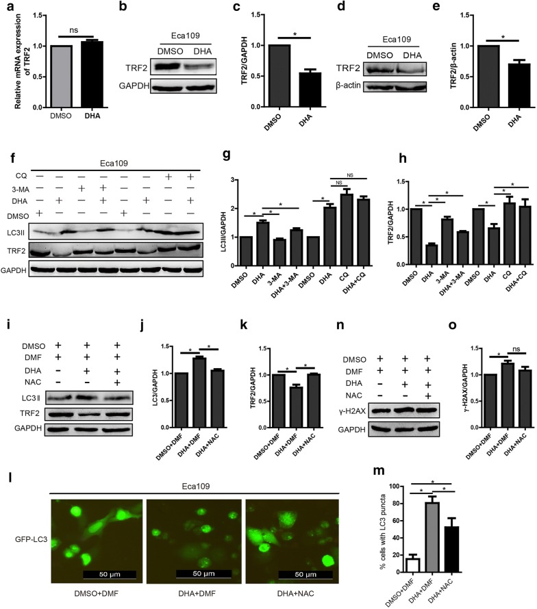
DHA downregulates the expression of TRF2 and the mechanisms. a–c The mRNA (a) and protein (b) expression of TRF2 in cells treated with DHA (100 μM) and the statistical chart (c). d, e The expression of TRF2 protein in tumor tissues from the xenograft mouse model treated with DHA (100 μM) or DMSO (d) and the statistical chart (e). f–h The expression of LC-3 and TRF2 in DHA (100 μM) treated cells after autophagy was inhibited by 3-MA (10 mM) or CQ (20 μM) (f) and the statistical chart (g, h). (i–k) The expression of LC-3 and TRF2 in DHA (100 μM) treated cells after ROS was blocked by NAC (5 mM) (i) and the statistical chart (j, k). l, m GFP-LC3 expression and puncta formation in DHA (100 μM) treated cells (l) and the statistical chart (m). n, o The expression of γ-H2AX in DHA (100 μM) treated cells after ROS was blocked by NAC (5 mM) (n) and the statistical chart (o). Data represent mean ± S.D. *p < 0.05 was significant difference between DHA-treated and control group

