FIGURE 2:
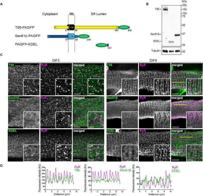
T95, Sec61β, and KDEL localize, respectively, in SR membranes and lumen. (A) Schematic representation of T95, Sec61β, and KDEL constructs fused to PAGFP, with N-terminal (N), C-terminal (C), and transmembrane (TM) domains sequence length and localization regarding the SR membrane (cytoplasm, membrane [Mb] and SR lumen). T95-PAGFP: TM in black, N and C-terminal in yellow and PAGFP in green; Sec61β: TM, N-, and C-terminal in blue and PAGFP in green; PAGFP-KDEL: PAGFP in green and KDEL in purple. Amino acid numbers from N-terminal to C-terminal end are indicated. (B) Western blot analysis has been performed with antibody against GFP on 10 μg triadin KO cells lysates transduced with T95-PAGFP or PAGFP-KDEL or Sec61β-PAGFP. Three bands are detected at 150 kDa (T95-PAGFP), 37 kDa (Sec61β-PAGFP), and 28 kDa (PAGFP-KDEL). The β-tubulin has been used as a loading control (bottom panel). (C) DIF3 (left) and DIF9 (right) myotubes expressing T95, Sec61β, or KDEL immunolabeled with anti-GFP (green) and anti-RyR1 (magenta) antibodies. Scale bars = 5 μm. Insets of T95, Sec61β, or KDEL immunolabelings with that of RyR1 are shown. Scale bars = 2 μm. Percentages of colocalization are shown on Figure 6D and Supplemental Figure S7 for DIF9 and DIF3, respectively .(D) Plot profiles of constructs (green) vs. RyR1 (magenta) intensities as a function of distance for 10 μm (five successive triads as shown by the yellow line on C) in TRDN KO myotubes at DIF9.

