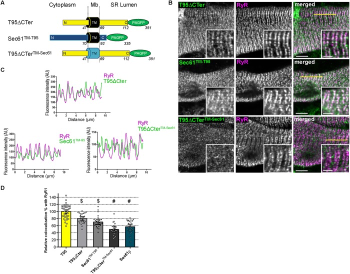
FIGURE 6:
Triadin TM domain is necessary for localization at triads. (A) Schematic representation of T95 ΔCter, Sec61 TM-T95, and T95ΔCTer TM-Sec61 constructs fused to PAGFP with N-terminal (N), C-terminal (C), and transmembrane (TM) domains sequence length and localization regarding the SR membrane (cytoplasm, membrane [Mb] and SR lumen). T95ΔCTer-PAGFP: TM in black, N, and C-terminal in yellow and PAGFP in green; Sec61TM-T95: TM in black, N- and C-terminal in blue, and PAGFP in green; T95ΔCTerTM-Sec61: TM in blue, N- and C-terminal in yellow, and PAGFP in green. Amino acid numbers from N-terminal to C-terminal end are indicated. (B) DIF9 myotubes expressing T95ΔCTer, Sec61TM-T95, and T95ΔCTer TM-Sec61 immunolabeled with anti-GFP (green) and anti-RyR1 (magenta) antibodies. Scale bars = 5 μm. Insets of T95 ΔCTer, Sec61TM-T95, or T95ΔCTerTM-Sec61 immunolabelings with that of RyR1 are shown. Scale bars = 2 μm. (C) Plot profiles of constructs (green) vs. RyR1 (magenta) intensities as a function of distance for 10 μm (five successive triads as shown by the yellow line in B) in TRDN KO myotubes at DIF9. (D) Percentage of PAGFP constructs colocalized with RyR1 labeling and normalized to that of DIF9 T95. Values are means ± c.i.; n = 40 cells from four independent experiments for T95; n = 26 cells from three independent experiments for Sec61β; n = 28 cells from three independent experiments for T 9 5 ΔCter; n = 21 cells from two independent experiments for T9 5 ΔCter TM-Sec61; n = 27 cells from two independent experiments for Sec61TM-T95. Using a parametric one-way analysis of variance followed by Tukey post-hoc comparisons: $, comparison with Sec61β: T95ΔCter (p < 0.0001) and Sec61TM-T95 (p < 0.05); #, comparison with T95ΔCter (p < 0.0001).

