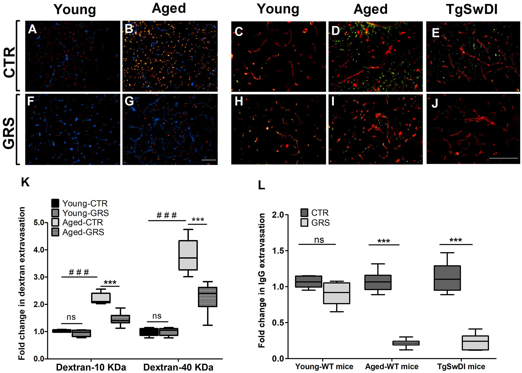
Figure 1. Granisetron enhanced BBB tightness in mice brains.

Mice were treated with granisetron (3 mg/day) for 28 days. (A,B,F,G) Representative brain sections measured using two different molecular weight fluorescents tagged dextran 10KD (red) 40 KD (green) as exogenous markers for BBB permeability and anti-Collagen IV antibody (Blue) to detect microvessels where (A) young control group, (F) young granisetron group, (B) aged control group, and (G) aged granisetron group. (K) Optical density semi-quantification of dextran 10 KD and 40 KD extravasation. (C-E, H-J) Representative brain sections stained with anti-mouse IgG antibody to detect IgG extravasation (green) and anti-collagen antibody (red) in wild type and TgSWDI mice brains and IgG levels quantification where (C) young control group, (H) young granisetron group, (D) aged control group, (I) aged granisetron group, (E) control TgSwDI group, (J) granisetron TgSwDI group, (L) optical density semi-quantification of IgG extravasation. All values in K were normalized to Young-CTR (1.0). GRS treatment values in L were normalized for each group corresponding CTR (1.0). Data are presented as box-and-whisker plots representing median and interquartile range (IQR), with minimum and maximum values. CTR is for control group i.e. vehicle-treated mice, GRS for granisetron, ns = not significant, ***P < 0.001, ###P<0.001 (Students t-test, n=5–7 mice). Scale bar, 200 μm.
