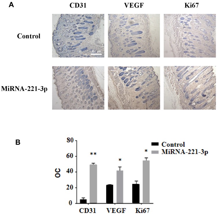
Figure 8.
Immunohistochemical staining of miRNA-221-3p–treated skin wound tissue in diabetic mice. Representative immunohistochemical images (A) and summary data (B) showing immunostaining in the skin wound tissue from diabetic mice treated with miRNA-221-3p (expressed as integrated optical density/total area; OC) (magnification, 200×). All values are presented as means and standard deviations (n = 3). Statistically significant differences are indicated by *P < 0.05 and **P < 0.01, compared with the control group treated with PBS.

