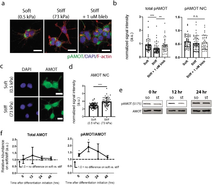
FIGURE 3:
Stiffness regulates AMOT phosphorylation and localization. (a) Representative immunofluorescence images of DAPI (blue), F-actin (red), and endogenous pAMOT (green) in NSCs after 24 h of differentiation on soft, stiff, or stiff + 1 µM blebbistatin culture conditions. (b) Quantification of total pAMOT intensity and nuclear/cytoplasmic localization ratio after 24 h of differentiation in various conditions. **p < 0.005, ***p < 0.001 by one-way ANOVA followed by Tukey’s post-hoc test, n.s. = not significant (p > 0.05). (c) Representative immunofluorescence images of AMOT (green) in NSCs after 24 h of differentiation on soft (0.5 kPa) or stiff (73 kPa) substrates (blue = DAPI). (d) Quantification of AMOT nuclear/cytoplasmic localization measured after 24 h of differentiation. Error bars represent SD (n = 54 cells for soft, n = 53 cells for stiff). ****p < 0.0001 by unpaired t test. (e) Representative Western blots and quantification of pAMOT/AMOT protein levels in cells differentiated for 0, 12, and 24 h of differentiation on soft vs. stiff substrates. (f) pAMOT and AMOT Western blot band intensities were normalized to GAPDH and β-actin, and the pAMOT/AMOT ratio of the normalized values on soft vs. stiff was calculated for each trial (n = 3 gels per timepoint). *p < 0.05 by one-sample t test against hypothetical value of 1.0.

