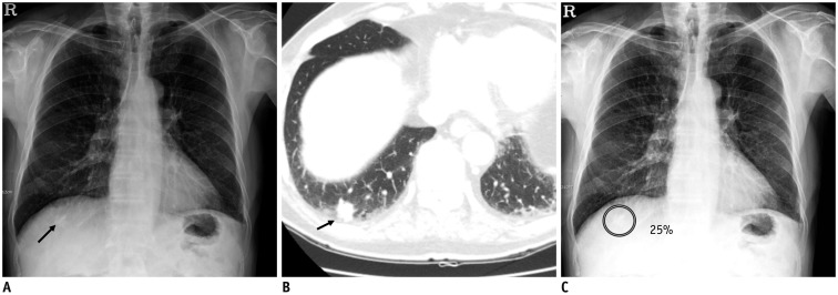
Fig. 1. Detection of lung nodules on chest X-ray.
A. Chest X-ray image shows nodular opacity at juxta-diaphragmatic right basal lung (arrow). B. Corresponding CT image shows 1.5-cm solid nodule at right lower lobe of lung (arrow). C. DL algorithm successfully detected nodule with output probability score of 25% (Courtesy of authors, DL algorithm is same as that in study by Nam et al. (30)). CT = computed tomography, DL = deep learning

