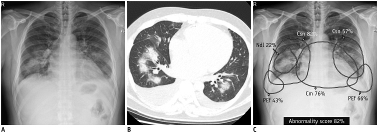
Fig. 2. Detection and differentiation of different abnormalities on chest X-ray.
Chest X-ray (A) and CT (B) obtained on same day from patient with pulmonary edema shows consolidation in both lung fields, bilateral pleural effusion, and mild cardiomegaly. DL algorithm classified chest X-ray image as abnormal, with 82% probability score. C. Algorithm identified Csns, PEf, and Cm on chest X-ray and localized each abnormality separately. Notably, algorithm recognized focal area of dense consolidation in right lower lung field as nodule (Courtesy of authors, DL algorithm is same as that in study by Kim et al. (132)). Cm = cardiomegaly, Csn = consolidation, Ndl = nodule, PEf = pleural effusion

