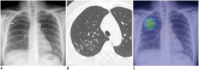
Fig. 3. Identification of chest X-ray with active pulmonary tuberculosis.
A. Chest X-ray of patient with active pulmonary tuberculosis shows subtle nodular infiltration in right upper lung field (arrow). B. Corresponding CT image shows clustered centrilobular nodules and mild bronchiectasis in right upper lobe of lung (arrow). C. DL algorithm successfully detected lesion, with heat map overlaid on chest X-ray (Courtesy of authors, DL algorithm is same as that in study by Hwang et al. (28)).

