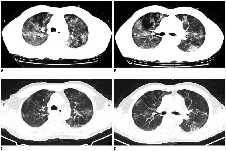
Fig. 4. CT findings on 24th and 29th day post-admission.
A, B. On day 24, consolidation was clearly absorbed. GGO regions were enlarged but GGO density had decreased. Some GGO was mixed with small patch consolidation. C, D. On day 29, GGO and small patch consolidation continued to absorb. Initial GGO on first CT scan and initial GGO and consolidation on second CT scan in apicoposterior segment of left upper lobe were clearly absorbed.

