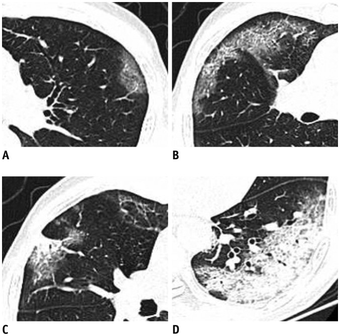
Fig. 1. Different presentations of lesions in both lungs of patient.
A. Pure GGO in left upper lobe. B. GGO with interlobular septal thickening in both right upper and middle lobe.
C. GGO with consolidation in right upper lobe. D. Mostly consolidation lesions in left inferior lobe. GGO = ground-glass opacity

