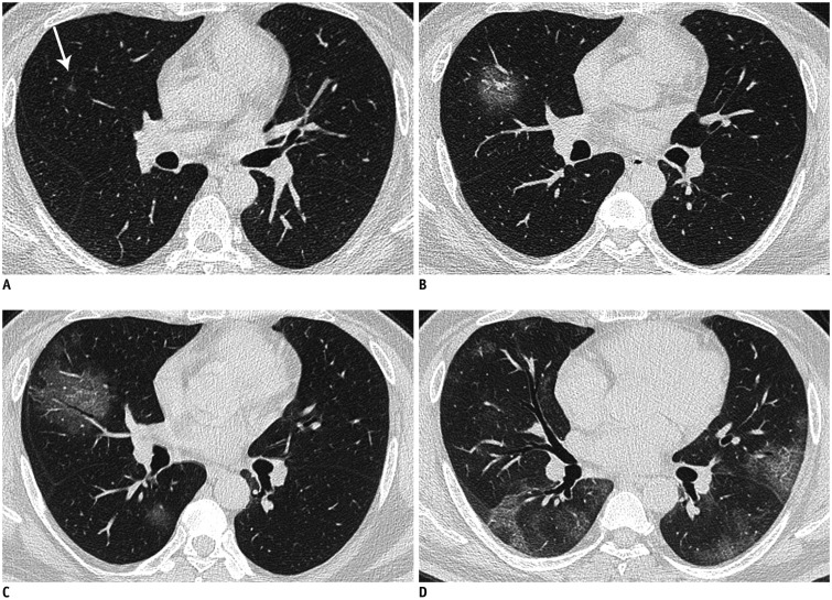
Fig. 3. Chest CT images of 48-year-old man with COVID-19 pneumonia.
A. Baseline CT image (2 days before onset of symptoms) shows suspicious solitary sub-centimeter ground-glass nodule 5 mm in size (arrow) in right middle lobe. Day 2 follow-up CT image (B) shows that vessels passing through lesion are thickened and air bronchogram also appears in lesion. Day 4 follow-up CT image (C) shows area of lesion is further enlarged. Moreover, similar ground-glass opacities also appear around initial lesion and in right lower lobe. Day 27 follow-up CT image (D) shows diffuse parenchymal abnormalities in periphery of whole lungs.

