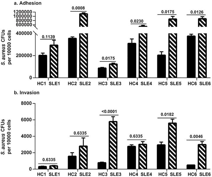
Fig 5. S. aureus demonstrated greater adherence to SLE keratinocytes compared to HC keratinocytes.
Keratinocytes from non-lesional skin from SLE patients and matched healthy controls (HC) were grown to confluence and exposed for 90 min to washed log phase S. aureus AH1263. S. aureus adhesion (A) and invasion (B) assays were performed as described in methods, and results are presented as S. aureus CFUs recovered per 10,000 keratinocytes. Results are presented as means from experiments performed at least in triplicate from each patient and paired control ± SEM (SLE n=6, HC n=6). Student’s t test was performed on each matched pair and the p values are denoted.

