Figure 2:
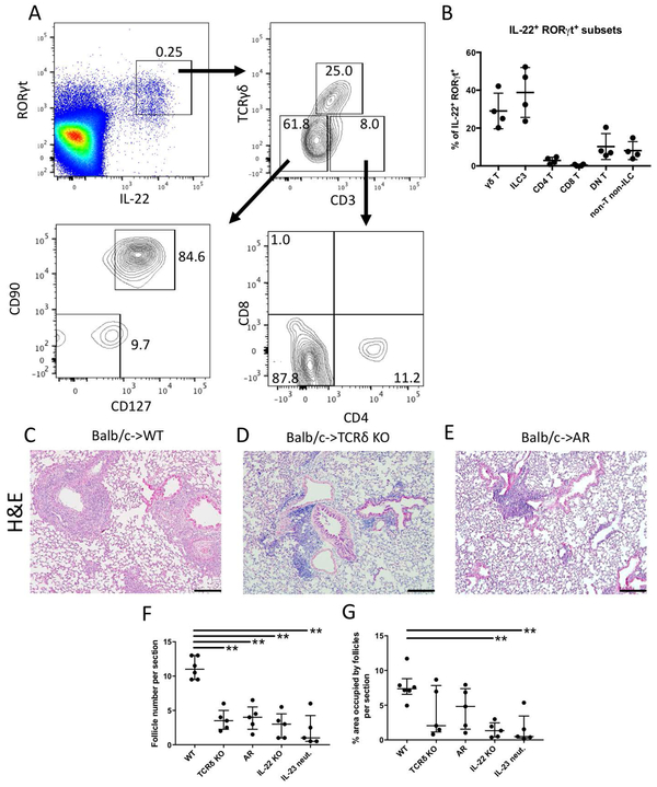
Graft-infiltrating γδ T cells and ILC3s are important sources of IL-22 after lung transplantation. (A) Flow cytometric characterization of IL-22+ cells types in tolerant Balb/c → B6 CD45.1 lung grafts reveals γδ T cells (RORγt+CD3+TCRγδ+) and ILC3s (RORγt+CD3−TCRγδ−CD127+CD90+) as main producers. Other IL-22+ cells include CD4+ T cells (RORγt+CD3+TCRγδ−CD4+CD8−), double-negative (DN) T cells (RORγt+CD3+TCRγδ−CD4−CD8−), CD8+ T cells (RORγt+CD3+TCRγδ−CD4−CD8+) and non-T non-ILCs (RORγt+CD3−TCRγδ−CD127−CD90−). Plots are gated on live single CD45.1+ cells. (B) Quantification of RORγt+IL-22+ cell subpopulations in tolerant lung grafts based on the gating plots described above. Histological appearance of lung allografts 30 days after transplantation into (C) wildtype (WT), (D) γδ T cell-deficient (TCRδ KO) recipients and (E) AR mice (RORγt-cre Ahrfl/fl) that received peri-operative co-stimulatory blockade. Quantification of the (F) number of lymphoid follicles and (G) percentage of total area occupied by lymphoid follicles per section 30 days after transplantation into various recipients. n≥4 for all conditions. **p<0.01. IL-22 KO: IL-22-deficient; IL-23 neut: IL-23 neutralization. Scale bars: 100μM.

