Figure 5:
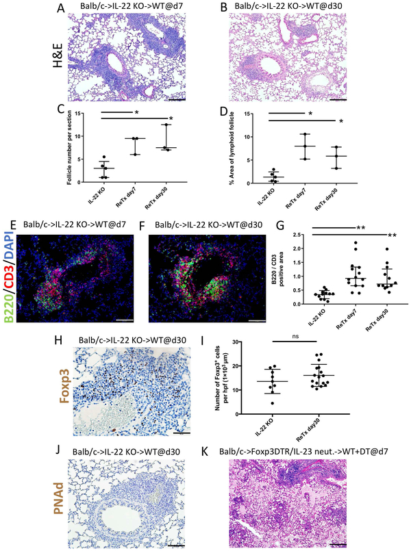
Late reconstitution of IL-22 expression mediates B cell recruitment to lymphoid follicles. Histological appearance of Balb/c lung grafts initially transplanted into IL-22 deficient recipients that received peri-operative costimulatory blockade and then retransplanted into non-immunosuppressed wildtype secondary recipients 30 days later. Images show lung allografts harvested (A) 7 and (B) 30 days after retransplantation. n=3. Scale bars =100 μm. Quantification of lymphoid follicle (C) number and (D) percent of total area per section in IL-22-deficient recipients 30 days after primary transplantation, as well as 7 and 30 days after retransplantation into wildtype secondary hosts. Histological appearance of B cell (green), T cell (red), and DAPI (blue) immunofluorescent staining from Balb/c lung grafts (E) 7 and (F) 30 days after retransplantation (scale bars =50 μm) and (G) quantification of B220 / CD3 cell ratio per lymphoid follicle among the various conditions. Each dot represents one lymphoid follicle (n≥2 mice per group). Immunostaining for Foxp3 (brown) (H) 30 days after retransplantation and (I) quantification of Foxp3 cells per lymphoid follicle at 30 days among various conditions. Scale bars =50 μm. Each dot represents one lymphoid follicle (n≥2 mice per group). Immunostaining for PNAd (brown) (J) 30 days after retransplantation. Scale bars =50 μm. (K) Histological appearance of Balb/c lung grafts initially transplanted into IL-23 neutralized B6 Foxp3-DTR recipients that received peri-operative costimulatory blockade and then retransplanted into non-immunosuppressed wildtype B6 recipients 30 days later that received DT at the time of retransplantation. Images are shown 7 days after retransplantation (H&E) (n=4). Scale bars =100 μm. ns= not significant; *p<0.05; **p<0.01.

