
Figure 1.
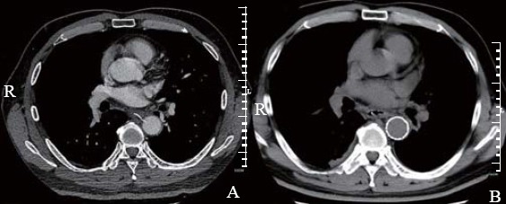
Computed tomographic images of case 1 before and after TEVAR. A: a sharp pin-like foreign body perforated left wall of esophagus and penetrated the right side of aorta on the plane of the 7th thoracic vertebra; B: a stent sealed the same site where the EFB had been.