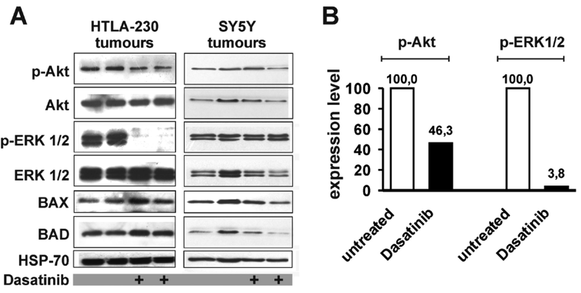
Figure 5.

A: western blot analyses to detect phospho-Akt (p-Akt), Akt, phospho-Erk1/2 (p-Erk1/2) and Erk1/2, BAX and BAD in cell extracts from tumours of untreated and Dasatinib-treated animals transplanted with HTLA-230 (blots on the left) or with SY5Y cells (blots on the right). Protein amount loaded in each lane was normalized by HSP-70 levels. B: Densitometric analysis of phospho-Akt and phospho-Erk1/2 in untreated and Dasatinib-treated animals injected with HTLA-230 cells. Each densitometric value represents the mean of 2 untreated and 2 Dasatinib-treated samples from blot in panel A. Values were normalized for the levels of total Akt and Erk1/2. Expression levels in untreated samples were arbitrarily set to 100.
