
Figure 8.
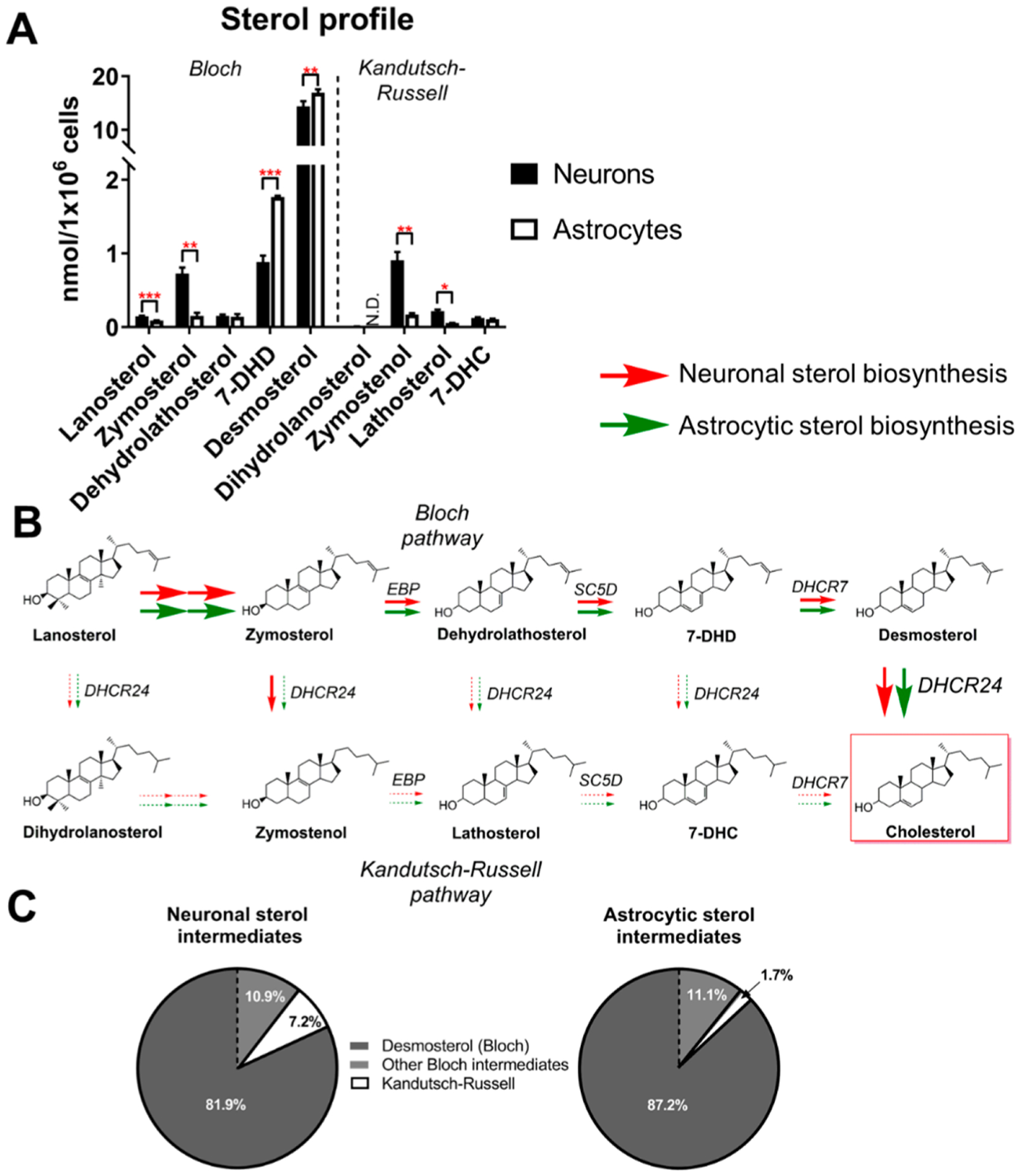
Both neurons and astrocytes synthesize cholesterol via the Bloch pathway. (A) GC-MS profile of postlanosterol cholesterol biosynthesis intermediates in cultured neurons and astrocytes. Cells were cultured in a defined cholesterol-free medium. Intermediates in the Bloch pathway are shown on the left, while those from the Kandutsch-Russell pathway are depicted on the right of the graph. Sterol levels were determined at DIC6 and values correspond to mean ± SEM of 3 replicates. Statistical significance is denoted by asterisks (*p < 0.05, **p < 0.01, ***p < 0.001, ND − nondetectable). (B) Schematic representation depicting the preferential routes for the conversion of lanosterol into cholesterol by cultured neurons and astrocytes. Red and green arrows represent the pathways utilized by neurons and astrocytes, respectively. Arrow thickness corresponds to the strength of pathway utilization. (C) Pie chart representation denoting the percent of Bloch and Kandutsch-Russell intermediates in cultured neurons and astrocytes at DIC6. Note that desmosterol is the most abundant intermediate in both neurons and astrocytes and that both cell types have higher levels of intermediates from the Bloch pathway, suggesting that the Kandutsch-Russell pathway utilization is miniscule at this developmental stage.