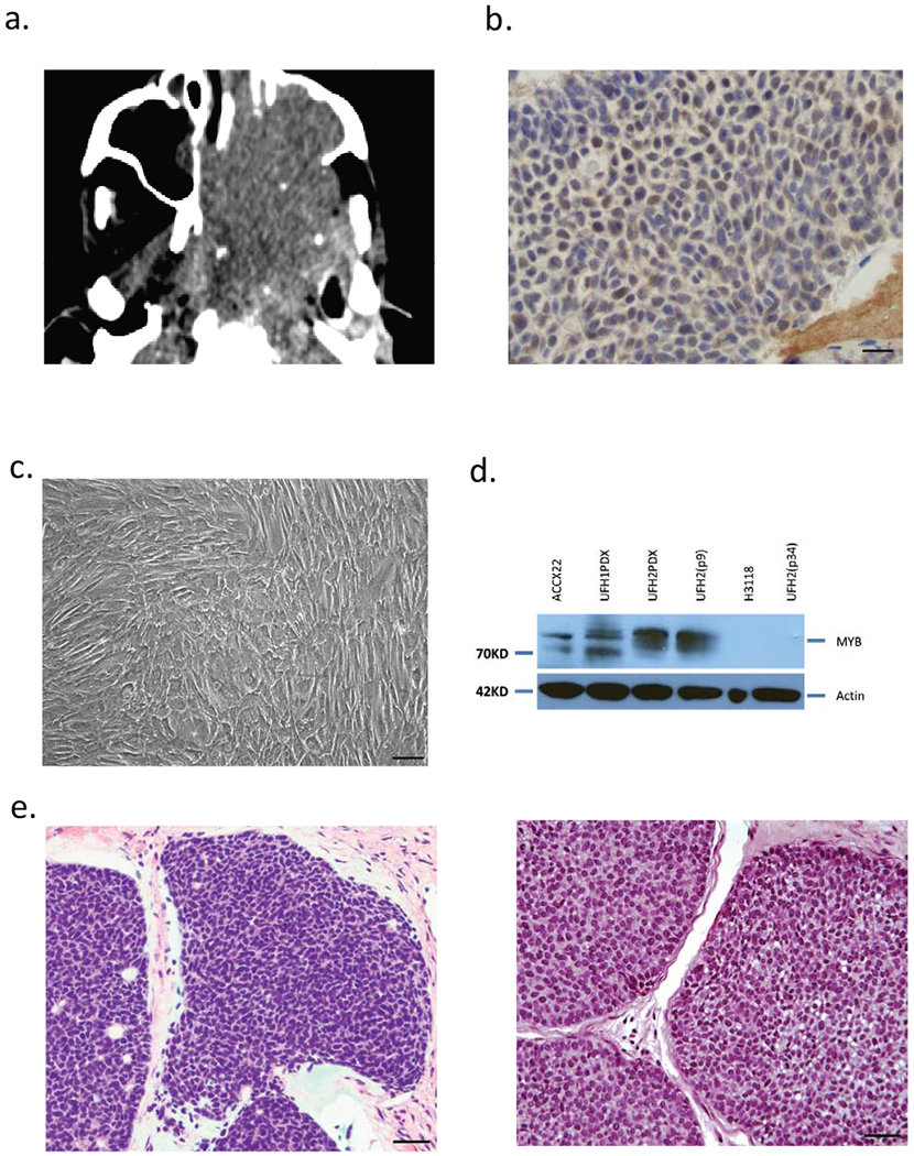
Figure 1.

Establishment of human ACC PDX and cell line (UFH2). (a) CT image of unresectable ACC requiring palliative debulking surgery. (b) Nuclear anti-MYB immunohistochemical staining in tumor biopsy. (c) UFH2 tumor cells in vitro at confluence. (d) MYB protein immunoblot (Anti-Myb, abcam, #ab45150) using ACCX22 (positive control), H3118 mucoepidermoid (negative control) and UFH1 PDX and UFH2 PDX and cell line at indicated passage number. (e) H&E staining primary ACC tumor biopsy section (left) and excised matched UFH2 PDX xenograft section (right).
