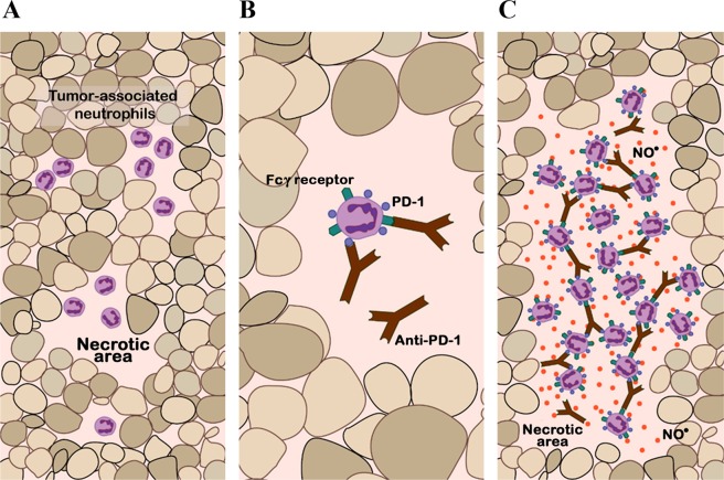
Figure 8.
Schematic representation of the possible mechanism of action of neutrophils. (A) Basal situation of a patient-derived xenograft. Necrotic areas are represented in a squamous lung cancer, in which an immune infiltrate of murine myeloid cells, including neutrophils, is evident. (B) Intra-tumor distribution of anti-PD-1 therapy. The anti-PD-1 antibody can bind to the PD-1 receptor on the neutrophil membrane; in addition, anti-PD-1 can recognize fragment crystallizable (Fc) gamma receptors (FcγR) and binds to their antibody Fc region. (C) Acute inflammatory reaction and neutrophil-mediated opsonization. Following on from (B), there is an inflammatory chain reaction and neutrophils acquire cytotoxic capacity and release nitric oxide (red circles in the figure), resulting in more extensive necrotic areas and subsequent tumor regression.

