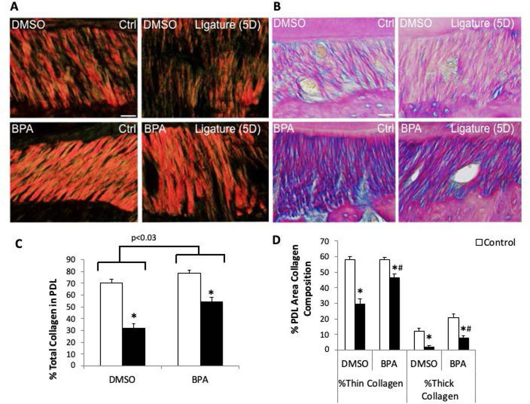
Figure 3:
Inhibition of TG activity in vivo restores PDL collagen following 5-day (5D) ligature. A). Representative polarized PSR-stained PDL sections of mice with and without 5-day ligature with vehicle only (DMSO) or vehicle with 5-(Biotinamido) pentylamine (BPA) injections. B). Representative Herovici-stained PDL images. Pink/red = thick/mature collagen fibers, blue/purple = thin/young collagen, yellow = cell death, black = nuclei. Each image oriented with tooth on top, PDL centered, and alveolar bone on bottom. Images shown are of PDL surrounding second maxillary molar. Scale bars = 25 μm. n = 5 mice per group. C). PSR quantification of PDL collagen content following 5-day ligature +/− DMSO/BC injections. D). PSR quantification of the percent thick versus thin collagen fiber area in PDL following 5-day ligature +/− BPA injection. *p < 0.05 between control and ligature, #p < 0.05 between DMSO and BPA injections, as determined by Student’s t test. n = 5 mice per group.

