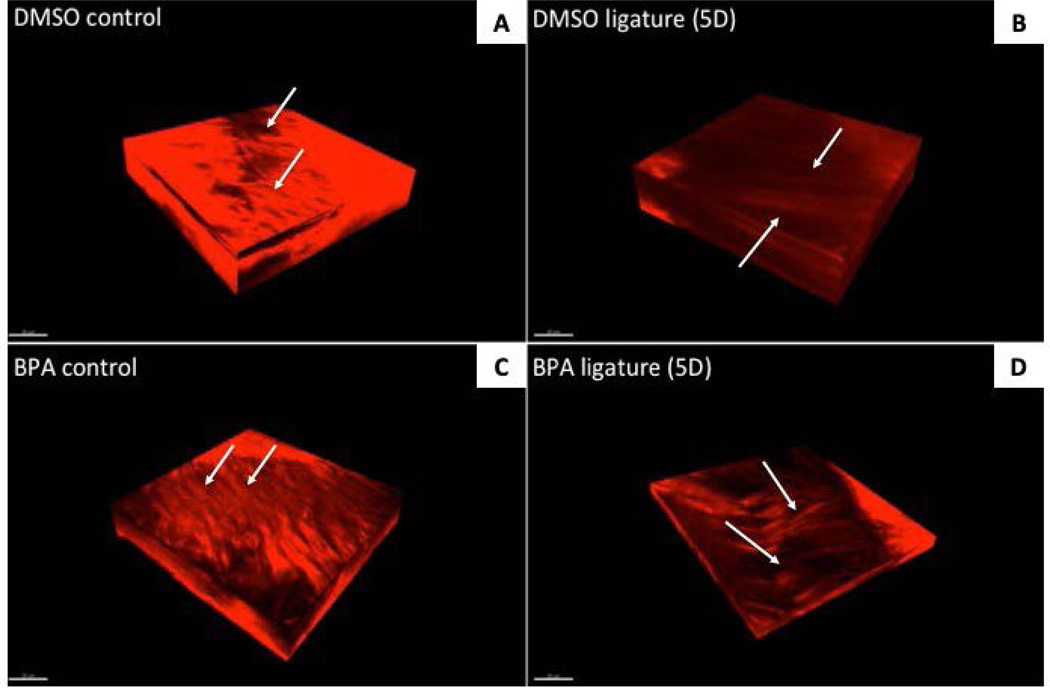
Figure 4:
Visualization of native collagen fiber morphology by second harmonic generation (SHG) in PDL treated with inhibitors of TG activity in 5D ligature mice. SHG orthogonal volume images generated from z-stacks taken at 0.2μm/slice for depths of up to 60μm of native PDL +/− DMSO/BPA injections following 5-day ligature treatment. Apparent increases in collagen fiber morphology and organization are observable in BPA-treated PDL (panels C & D) versus those treated with DMSO (A and B). The appearance of thick collagen fiber bundles is apparent in 5D BPA-treated PDL (D) versus vehicle only (B). Arrows, collagen fiber bundles. Scale bar = 20μm.

