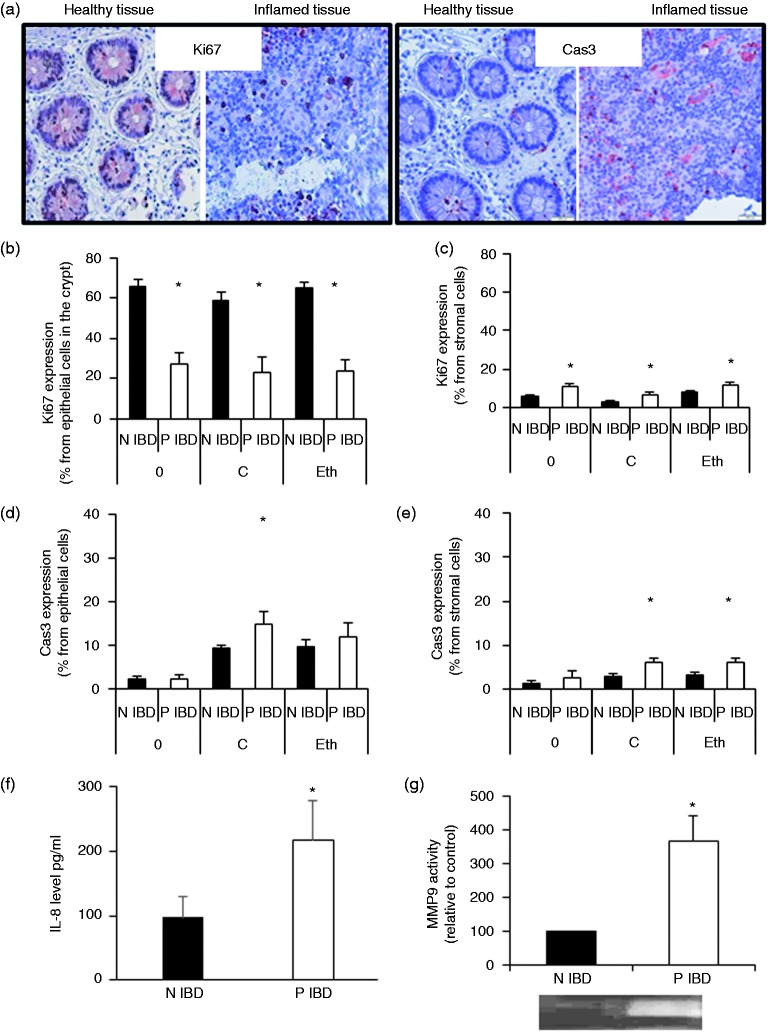
Figure 2.
Cultured biopsies from inflamed colon areas retain characteristics similar to those found in vivo.
Biopsies were taken during endoscopy from inflamed and uninflamed areas of inflammatory bowel disease patients. For each immunohistochemical analysis two biopsies (N and P) were immediately fixed in formaldehyde (time 0) for Ki67 expression (proliferation) and cleaved caspase 3 expression (apoptosis), and the rest of the biopsies were cultured with (one N and one P)/without (one N and one P) ethanol for 6 hours, and then analyzed for stromal/epithelial cell Ki67 and cleaved caspase 3 expression. Moreover, secretomes collected from P and N (N inflammatory bowel disease) cultured biopsies were analyzed for matrix metalloproteinase 9 activity and interleukin-8 levels. Presented are representative photomicrographs of inflamed/non-inflamed biopsies of inflammatory bowel disease patients stained for Ki67 and cleaved caspase 3 (a), graphs that show Ki67 and cleaved caspase 3 expression in epithelial ((b) and (d)) and stromal ((c) and (e)) cells, and graphs that show interleukin-8 levels (f) and matrix metalloproteinase 9 activity (g) in the secretomes. (b) to (e) Time 0 and biopsies cultured for 6 hours in medium: n = 8–9 biopsies (16–18 pictures) and biopsies cultured for 6 hours with ethanol in the medium: n = 5–8 patients (10–16 pictures); (f) n = 8; (g) n = 5. *indicates statistically significant results (p < 0.05).
0: time 0; C: biopsies cultured for 6 hours in medium; Eth: biopsies cultured for 6 hours with ethanol in the medium; IBD: inflammatory bowel disease; IL-8: interleukin-8; MMP9: matrix metalloproteinase 9; N: uninflamed; P: inflamed.

