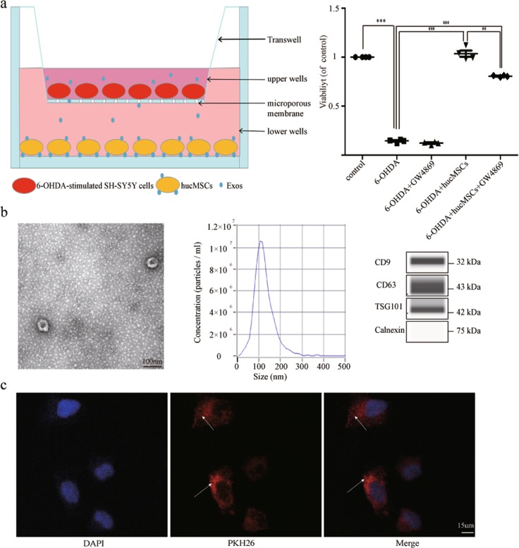
Fig. 3. The function and typical characteristics of Exos.
a The transwell system. hucMSCs and 6-OHDA-stimulated SH-SY5Y cells were co-cultured in a transwell system. GW4869 was used at a concentration of 10 µM to reduce the release of Exos from hucMSCs. After co-culturing for 8 h, the viability of 6-OHDA-stimulated SH-SY5Y cells was determined (n = 4). b Representative transmission electron microscopy (TEM) images of Exos. Scale bar, 100 nm. Size distributions of Exos were measured using nanoparticle tracking analysis (NTA). Western blotting analysis of Exos markers including CD9, CD63, Tsg101, and Calnexin. c PKH26-labeled Exos (red) and DAPI (blue). Arrows indicate Exos. All data represent mean values ± SD. *P < 0.05; **P < 0.01; ***P < 0.001.

