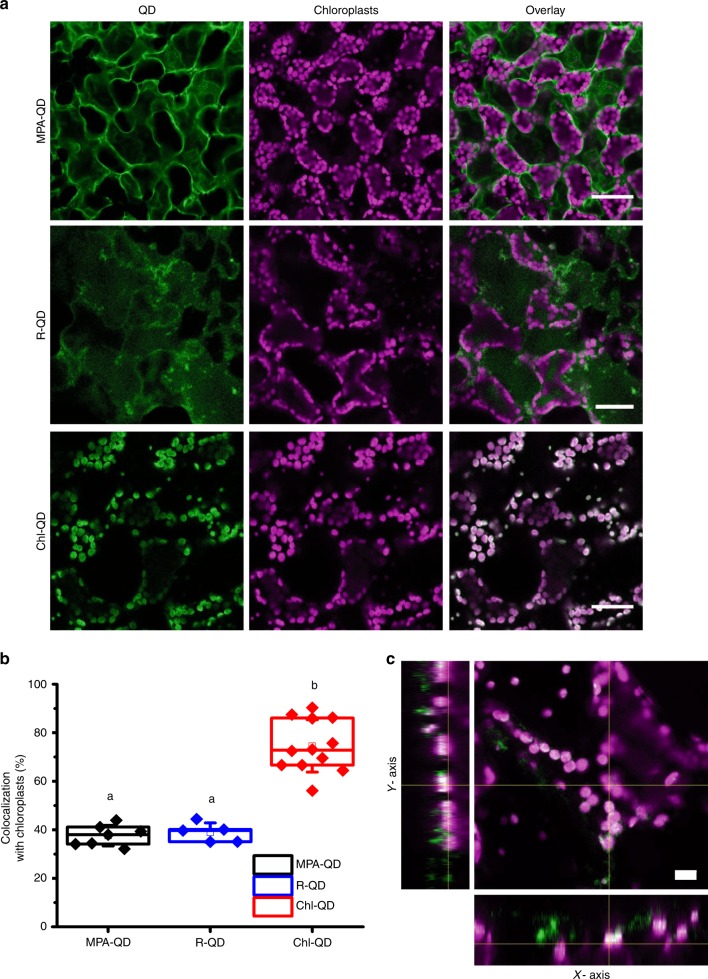
Fig. 3. Targeted delivery of quantum dots to chloroplasts of Arabidopsis leaf mesophyll cells.
a Confocal microscopy images of chloroplasts in leaf mesophyll cells indicating a higher degree of colocalization of QD coated with guiding peptide (Chl-QD, n = 12) with chloroplasts compared to QD without targeting peptide (MPA-QD, n = 7) and QD coated with a randomized amino acid sequence of the guiding peptide (R-QD, n = 5). Scale bar, 40 µm. b Colocalization rates of Chl-QD (n = 12) with chloroplasts compared to MPA-QD (n = 7) and R-QD (n = 5). Statistical comparisons were performed by one-way ANOVA based on Duncan’s multiple range test (two tailed). Lower case letters represent significance at P < 0.05. Box plot error bars represent standard deviation, boxes are the interquartile range from the first to the third quartile with squares as the medians, and horizontal line represents the mean. c Orthogonal views of different planes from confocal images (z-stack) of Chl-QD colocalization within chloroplasts. Scale bar, 10 µm.

