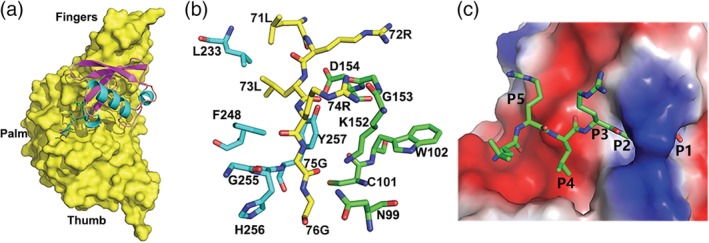
FIGURE 3.

Hypothetical model of the interaction of ubiquitin with the SADS‐CoV PLP2 active site based on the structure of SARS‐CoV PLpro bound with Ub. (a) The model of SADS‐CoV PLP2 bound with ubiquitin. A surface representation of the SADS‐CoV PLP2 is shown complexed with modeled ubiquitin. The C‐terminal of ubiquitin is shown by a ball‐and‐stick representation. (b) Modeled interactions between the C‐terminal tail of ubiquitin and the SADS‐CoV PLP2. Ubiquitin residues are colored in yellow carbon, while PLP2 residues are shown in cyan or green carbons. (c) A surface representation of the SADS‐CoV PLP2 with a tunnel of the active site bound by C‐terminal five residues of ubiquitin. The P1–P5 positions of ubiquitin are labeled
