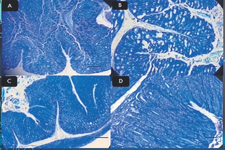
Fig. 2.
A Stomach epithelium of mice after treatment with L. rhamnosus stained with Giemsa stain. Original magnification, ×40. B Stomach epithelium of mice after treatment with L. acidophilus stained with Giemsa stain. Original magnification, ×40. C Stomach epithelium of mice after treatment with L. plantarum stained with Giemsa stain. Original magnification, ×40. D Stomach epithelium of mice after treatment with all three lactobacilli stained with Giemsa stain. Original magnification, ×40.

