
Figure 2. NONU-1 Contains a Conserved Smr Domain Required for Nonstop mRNA Decay.
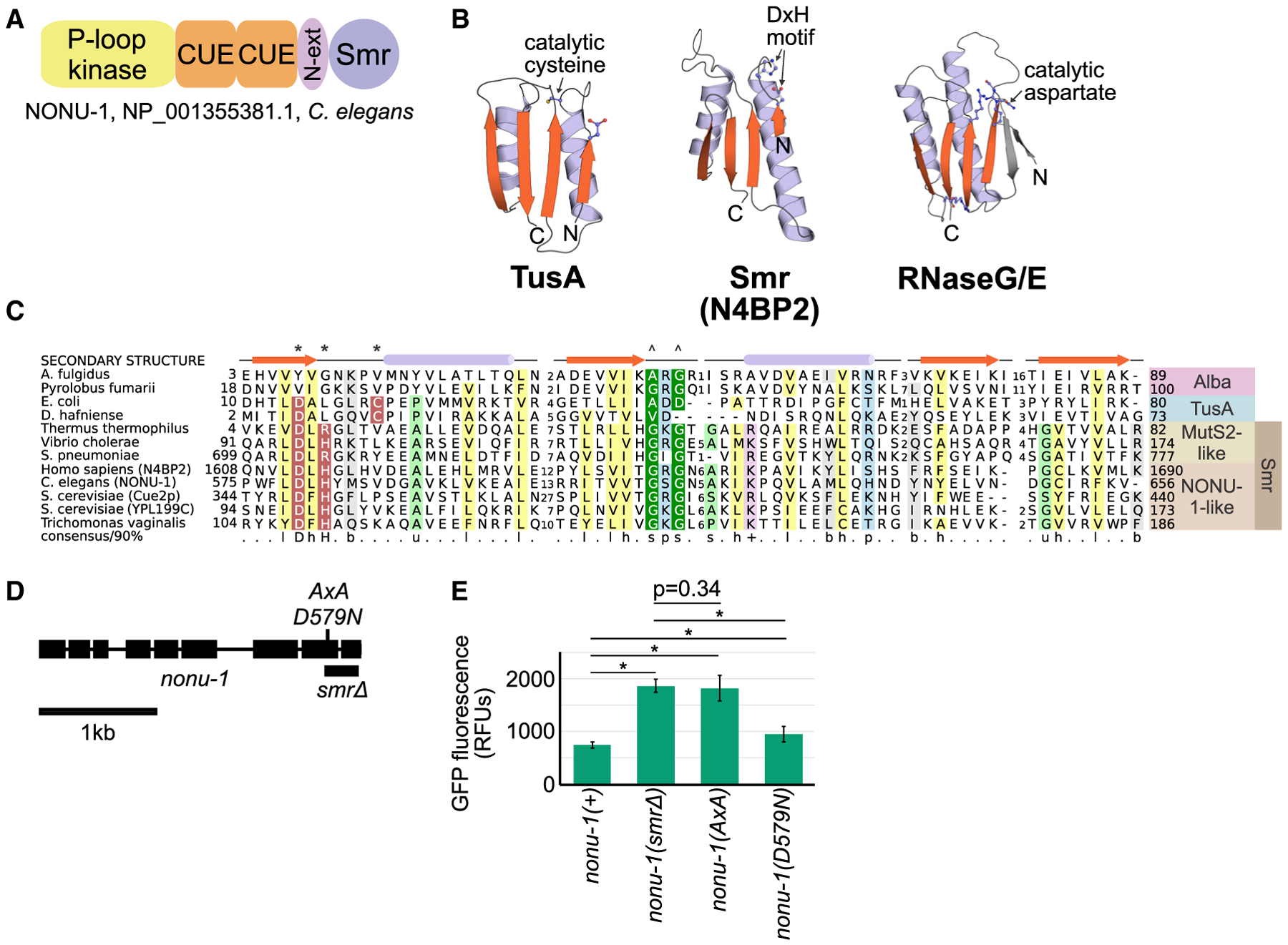
(A) Protein domain schematic of NONU-1. See text for details.
(B) Structures of representatives of the IF3-C fold domain, including the Smr domain from the human homolog of NONU-1, N4BP2. See also Figure S3C.
(C) Multiple sequence alignment of the TusA-Alba-Smr assemblage with protein secondary structure diagram and conserved amino acids highlighted. Asterisk and caret indicate amino acids involved in catalysis and substrate recognition, respectively, in TusA- or MutS2-like proteins. See also Figure S3A.
(D) Gene diagram showing the nonu-1 locus and mutations generated by CRISPR/Cas9.
(E) Quantification of GFP expression for the indicated strains (as in Figure 1E). p values from Student’s t test; *p < 10e-6. Compare with Figure 1E. See also Figure S3.