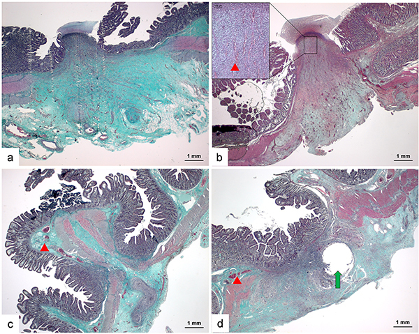
Figure 4. Architecture of the anastomoses. Panels a and b, Anastomoses established by LigaSure had gaps, which were filled by abundant collagen fibers (stained in green color in trichromatic staining images) between two extremities of the muscle layer of each side of the anastomosis and regenerative capillaries could be identified between the collagen fibers (red arrowhead). Panels c and d, Anastomoses established by GIA or suture had a shorter distance of gaps between two sides. Their muscle layers were connected mechanically by titanium staples or sutures. The pre-existing capillaries (red arrowhead) could be observed in sub-mucosa and signs of revascularization were rare (scale bar 1 mm).

